Tap image to enlarge이미지를 누르면 크게 볼 수 있습니다
Application & Purpose작품의 목적
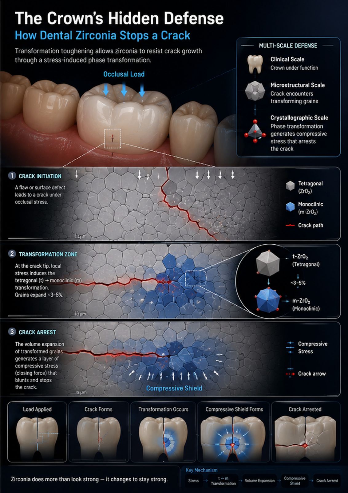
This scientific visualization explains transformation toughening in a dental zirconia crown. It links occlusal loading at the clinical scale to crack initiation, stress-induced tetragonal-to-monoclinic transformation, local volume expansion, compressive shielding, and crack arrest at the microstructural scale. The goal is to make a difficult fracture-mechanics concept immediately visible.
이 과학 시각화는 치과용 지르코니아 크라운의 변태 강화를 설명합니다. 임상 규모의 교합 하중을 균열 발생, 응력 유도 정방정→단사정 변태, 국소 부피 팽창, 압축 차폐, 그리고 미세구조 규모의 균열 정지까지 연결합니다. 어려운 파괴역학 개념을 한눈에 보이게 만드는 것이 목표입니다.
AI Tools & WorkflowAI 도구와 작업 흐름
ChatGPT image generation translated a materials-science storyboard into a multiscale illustration. I reviewed the crown geometry, grain structure, crack path, phase labels, and stress directions, separated the artwork, and exported it as an A3 single-page PDF.
ChatGPT 이미지 생성으로 재료과학 스토리보드를 다중 규모 일러스트로 옮겼습니다. 크라운 형상, 결정립 구조, 균열 경로, 상 라벨, 응력 방향을 검토하고 작품을 분리해 A3 단일 페이지 PDF로 내보냈습니다.
Process & Iterations제작 과정과 반복
I first reduced the mechanism to five causal steps: load, crack initiation, transformation-zone formation, compressive shielding, and crack arrest. A clinical crown alone did not reveal why zirconia behaves differently from conventional brittle ceramics, while an isolated grain diagram lacked dental context. I therefore combined three nested scales: the loaded crown, the polycrystalline crack path, and the tetragonal-to-monoclinic crystal change. The prompt emphasized a localized transformed zone near the crack tip rather than transformation throughout the crown. After generation, I checked the loading direction, crack continuity, placement of transformed grains, phase terminology, and the 3–5% volume-expansion annotation. I then reviewed the typography and prepared the panel for A3 printing.
먼저 메커니즘을 하중, 균열 발생, 변태 영역 형성, 압축 차폐, 균열 정지의 다섯 인과 단계로 줄였습니다. 임상 크라운만으로는 지르코니아가 일반 취성 세라믹과 왜 다른지 드러나지 않았고, 결정립 도식만으로는 치과적 맥락이 부족했습니다. 그래서 하중을 받는 크라운, 다결정 균열 경로, 정방정→단사정 결정 변화라는 세 개의 중첩된 규모를 결합했습니다. 프롬프트에서는 크라운 전체가 아닌 균열 끝 근처의 국소 변태 영역을 강조했습니다. 생성 후 하중 방향, 균열의 연속성, 변태된 결정립의 위치, 상 용어, 3~5% 부피 팽창 표기를 점검하고, 타이포그래피를 검토해 A3 인쇄용 패널로 준비했습니다.
A Researcher's Perspective연구자의 관점
AI made it possible to visualize clinical, microstructural, and crystallographic scales in one coherent field—something difficult to achieve with conventional figure assembly. The surprising strength was its ability to create an intuitive “defense zone” around the crack tip. The limitation was that plausible-looking grains and stress arrows are not automatically correct. Expert review remains essential because, in materials science, a small spatial or directional error can reverse the mechanism being communicated.
AI 덕분에 임상, 미세구조, 결정학적 규모를 하나의 일관된 화면에 담을 수 있었습니다. 기존 방식의 그림 조합으로는 어려운 일입니다. 놀라운 강점은 균열 끝 주변의 직관적인 '방어 영역'을 만들어낸 점이었습니다. 한계는 그럴듯해 보이는 결정립과 응력 화살표가 자동으로 정확하지는 않다는 것입니다. 재료과학에서는 작은 공간적·방향적 오류가 전달하려는 메커니즘을 뒤집을 수 있으므로 전문가 검토가 여전히 필수입니다.