Tap image to enlarge이미지를 누르면 크게 볼 수 있습니다
Application & Purpose작품의 목적
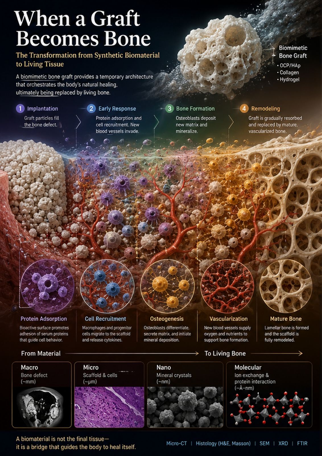
This infographic explains how a calcium-phosphate bone graft functions as a temporary scaffold rather than a permanent replacement. It follows implantation, protein adsorption, cellular recruitment, vascularization, osteogenesis, and remodeling across macro-to-molecular scales, helping non-specialists understand how an engineered material can guide the formation of living bone.
이 인포그래픽은 인산칼슘 골이식재가 영구적인 대체물이 아니라 임시 지지체로 작동하는 과정을 설명합니다. 이식, 단백질 흡착, 세포 모집, 혈관 형성, 골 형성, 재형성을 거시적 규모부터 분자 규모까지 따라가며, 공학적으로 설계된 재료가 어떻게 살아 있는 뼈의 형성을 이끄는지 비전문가도 이해할 수 있게 돕습니다.
AI Tools & WorkflowAI 도구와 작업 흐름
ChatGPT image generation was used to structure the scientific narrative and create the initial artwork. The composition was then reviewed for biological plausibility, separated into an A3 panel, typographically checked, and exported as a print-ready single-page PDF.
ChatGPT 이미지 생성으로 과학적 서사를 구성하고 초기 작품을 만들었습니다. 이후 생물학적 타당성을 검토하고, A3 패널로 분리한 뒤 타이포그래피를 점검해 인쇄용 단일 페이지 PDF로 내보냈습니다.
Process & Iterations제작 과정과 반복
I began with a stage-based storyboard derived from the biological sequence of graft-mediated bone regeneration. Early concepts separated the clinical defect, cell-scale events, and material chemistry too rigidly, so I redesigned them as one continuous landscape moving from graft particles to vascularized mature bone. The prompt specified OCP/HAp granules, a collagen-hydrogel matrix, protein adsorption, cell recruitment, osteoblast activity, vessel ingrowth, mineral deposition, and gradual graft resorption. After generation, I checked the order and spatial relationships of these events against my research experience, retained only scientifically interpretable elements, separated the panel from the paired draft, reviewed the labels and layout, and formatted it for A3 printing.
이식재를 통한 골 재생의 생물학적 순서를 바탕으로 단계별 스토리보드에서 출발했습니다. 초기 구상은 임상적 결손부, 세포 수준의 사건, 재료 화학을 너무 딱딱하게 분리해서, 이식재 입자에서 혈관화된 성숙 골까지 이어지는 하나의 연속된 풍경으로 다시 설계했습니다. 프롬프트에는 OCP/HAp 과립, 콜라겐 하이드로겔 기질, 단백질 흡착, 세포 모집, 조골세포 활동, 혈관 유입, 무기질 침착, 점진적 이식재 흡수를 명시했습니다. 생성 후에는 이 사건들의 순서와 공간 관계를 제 연구 경험과 대조해 확인하고, 과학적으로 해석 가능한 요소만 남긴 뒤 짝지어진 초안에서 패널을 분리하고 라벨과 레이아웃을 검토해 A3 인쇄용으로 정리했습니다.
A Researcher's Perspective연구자의 관점
AI was most useful for connecting events that are usually shown in separate figures: scaffold architecture, cell recruitment, vascularization, mineralization, and remodeling. Its strength was visual continuity; its limitation was scientific overconfidence, especially when inventing microscopic detail. A domain expert must therefore define the causal sequence, inspect every label and structure, and treat the generated image as an editable hypothesis—not as primary evidence.
AI는 보통 별개의 그림으로 나뉘어 표현되는 사건들, 즉 지지체 구조, 세포 모집, 혈관화, 무기질화, 재형성을 하나로 연결하는 데 가장 유용했습니다. 강점은 시각적 연속성이고, 한계는 특히 미세 구조를 지어낼 때 드러나는 과학적 과신입니다. 따라서 분야 전문가가 인과 순서를 정의하고 모든 라벨과 구조를 점검해야 하며, 생성된 이미지를 일차 증거가 아닌 편집 가능한 가설로 다루어야 합니다.