Tap image to enlarge이미지를 누르면 크게 볼 수 있습니다
Application & Purpose작품의 목적
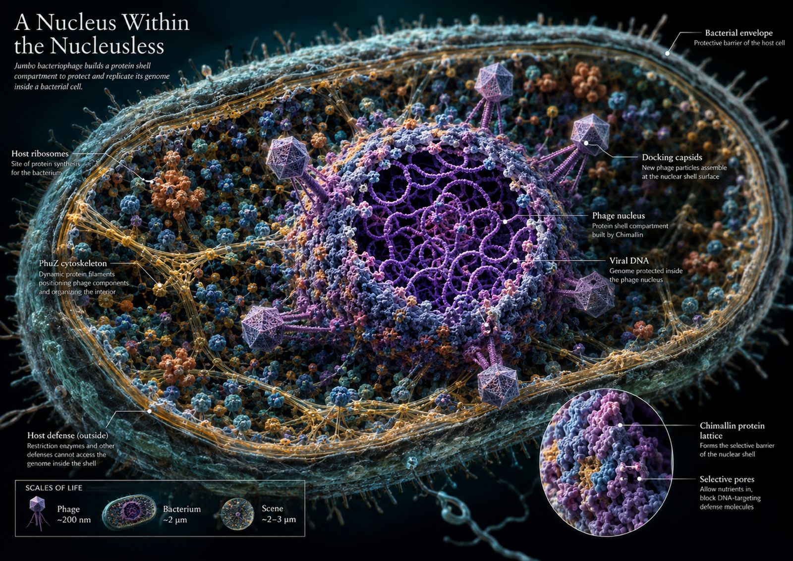
This artwork visualizes how jumbo bacteriophages construct a proteinaceous nucleus-like compartment inside bacteria. By revealing viral DNA protected within a chimallin shell, alongside host ribosomes, defense systems, and assembling capsids, it transforms an invisible cellular process into an accessible scene. The work aims to spark curiosity while communicating a surprising example of viral organization.
이 작품은 점보 박테리오파지가 세균 안에 단백질로 된 핵 유사 구획을 만드는 과정을 시각화합니다. 키말린 껍질 안에 보호된 바이러스 DNA를 숙주의 리보솜, 방어 체계, 조립 중인 캡시드와 함께 드러내어 보이지 않는 세포 과정을 접근 가능한 장면으로 바꿉니다. 호기심을 자극하는 동시에 바이러스 조직화의 놀라운 사례를 전달하는 것이 목표입니다.
AI Tools & WorkflowAI 도구와 작업 흐름
ChatGPT supported literature synthesis, concept development, prompt design, and scientific quality checks. OpenAI’s image model generated the artwork through iterative prompting. Python and ReportLab converted the final image into a single-page, print-ready A3 PDF at approximately 300 dpi.
ChatGPT가 문헌 종합, 개념 개발, 프롬프트 설계, 과학적 품질 점검을 도왔습니다. OpenAI 이미지 모델이 반복 프롬프팅으로 작품을 생성했습니다. 파이썬과 ReportLab으로 최종 이미지를 약 300dpi의 인쇄용 단일 페이지 A3 PDF로 변환했습니다.
Process & Iterations제작 과정과 반복
Early generations drifted toward a generic cell infographic and overfilled the scene with decorative molecular detail. I compared drafts against structural studies, then tightened prompts around the required relationships: viral DNA inside a chimallin-based compartment; host ribosomes and defense machinery outside; PhuZ organization and capsid assembly at the shell. I reduced explanatory clutter, tested alternative compositions, and selected the version with the clearest visual hierarchy at A3 scale. The final image was upscaled to 4961 × 3508 pixels, placed full bleed on an A3 landscape page, exported with ReportLab, and checked by rendering the PDF at print size.
초기 생성물은 일반적인 세포 인포그래픽으로 흘러갔고 장식적인 분자 세부로 장면이 가득 찼습니다. 초안을 구조 연구와 비교한 뒤 필요한 관계를 중심으로 프롬프트를 조였습니다. 키말린 기반 구획 안의 바이러스 DNA, 바깥의 숙주 리보솜과 방어 기구, 껍질에서의 PhuZ 조직화와 캡시드 조립입니다. 설명적 잡동사니를 줄이고 대안 구성을 시험한 뒤 A3 규모에서 시각 위계가 가장 명확한 버전을 골랐습니다. 최종 이미지는 4961×3508 픽셀로 업스케일해 A3 가로 페이지에 풀블리드로 배치하고 ReportLab으로 내보낸 뒤 인쇄 크기로 렌더링해 확인했습니다.
A Researcher's Perspective연구자의 관점
AI was most valuable as a rapid visual hypothesis generator: it revealed compositions I would not have sketched myself. Its limitation was equally clear. It often replaced unfamiliar biology with familiar visual clichés, such as membrane-like nuclei or fully assembled phages. Scientific accuracy therefore required active supervision, literature checking, and repeated constraint-setting. I would tell researchers to treat AI as a collaborative illustrator, not an authority: define the mechanism first, preserve failed iterations, and verify every visually persuasive detail.
AI는 빠른 시각 가설 생성기로서 가장 가치 있었습니다. 제가 직접 스케치하지 않았을 구성을 드러냈습니다. 한계도 그만큼 분명했습니다. 익숙하지 않은 생물학을 막 같은 핵이나 완전히 조립된 파지 같은 익숙한 시각적 클리셰로 자주 바꿔 놓았습니다. 그래서 과학적 정확성에는 적극적인 감독, 문헌 확인, 반복적인 제약 설정이 필요했습니다. 연구자들에게 말하고 싶습니다. AI를 권위자가 아닌 협업 일러스트레이터로 대하세요. 먼저 메커니즘을 정의하고, 실패한 반복을 보존하고, 시각적으로 설득력 있는 모든 세부를 검증하세요.